Amantadine can induce intra-epithelial deposition in the cornea
American Journal of Ophthalmology Case Reports Volume 19
Page 100852-
published_at 2020-08-05
アクセス数 : 294 件
ダウンロード数 : 81 件
今月のアクセス数 : 0 件
今月のダウンロード数 : 1 件
この文献の参照には次のURLをご利用ください : https://ir.lib.hiroshima-u.ac.jp/00051556
| File | |
| Title ( eng ) |
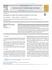
Amantadine can induce intra-epithelial deposition in the cornea
|
| Creator |
Yoshinaka Asayo
|
| Source Title |
American Journal of Ophthalmology Case Reports
|
| Volume | 19 |
| Start Page | 100852 |
| Abstract |
Purpose
Amantadine has been reported to cause various corneal complications, such as superficial punctate keratitis, corneal endothelial dysfunction, and corneal edema. However, there have been no reports of amantadine-induced deposits in the corneal epithelium. Here, we describe the first case with amantadine-induced deposits in the corneal epithelium proved by confocal biomicroscopy. Observations An 81-year-old woman presented with impaired vision in both eyes. She had been treated with amantadine for 9 years. Corrected visual acuity was 0.8 in both eyes. Furthermore, both eyes showed opacities in the corneal epithelial corneal layer. On confocal biomicroscopy, there were highly reflective deposits in corneal epithelial cells. There were no pathological findings in the stroma and endothelium. Two months after discontinuation of amantadine, corneal opacities disappeared, and visual acuity was 1.0 in both eyes. Conclusions Administration of amantadine can cause deposits in corneal epithelial cells. |
| Keywords |
Amantadine
Corneal opacity
Confocal microscopy
Corneal epithelial cell deposits
|
| Language |
eng
|
| Resource Type | journal article |
| Publisher |
Elsevier
|
| Date of Issued | 2020-08-05 |
| Rights |
© 2020 The Authors. Published by Elsevier Inc. This is an open access article under the CC BY-NC-ND license (http://creativecommons.org/licenses/by-nc-nd/4.0/).
|
| Publish Type | Version of Record |
| Access Rights | open access |
| Source Identifier |
[ISSN] 2451-9936
[DOI] 10.1016/j.ajoc.2020.100852
[DOI] https://doi.org/10.1016/j.ajoc.2020.100852
|