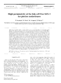
High permissivity of the fish cell line SSN-1 for piscine nodaviruses.
Diseases of Aquatic Organisms Volume 39 Issue 1
Page 37-47
published_at 1999-12-22
アクセス数 : 829 件
ダウンロード数 : 264 件
今月のアクセス数 : 2 件
今月のダウンロード数 : 2 件
この文献の参照には次のURLをご利用ください : https://ir.lib.hiroshima-u.ac.jp/00025659
| File | |
| Title ( eng ) |
High permissivity of the fish cell line SSN-1 for piscine nodaviruses.
|
| Creator |
Iwamoto Tokinori
Mori Koh-ichiro
Arimoto Misao
|
| Source Title |
Diseases of Aquatic Organisms
|
| Volume | 39 |
| Issue | 1 |
| Start Page | 37 |
| End Page | 47 |
| Abstract |
Seventeen isolates of piscine nodavirus from larvae or juveniles of 13 marine fish species affected with viral nervous necrosis (VNN) were examined for their infectivity to a fish cell line SSN-1. Based on cytopathic effects (CPE) and virus antigen detection by fluorescent antibody technique (FAT) after incubation at 25°C, the infectivity of these virus isolates was divided into 4 groups. Group 1, including 9 virus isolates from 4 species of grouper, 2 species of sea bass, barramundi, rock porgy, and Japanese flounder showed CPE characterized by rounded, granular cells with heavy cytoplasmic vacuoles within 3 d post-incubation (p.i.), and the monolayer partially or completely disintegrated over 3 to 6 d p.i. Scattered FAT-positive cells appeared at 3 h p.i. and spread through the cell sheet with an increasing fluorescence signal over 24 h p.i. Group 2, consisting of 3 virus isolates from striped jack, induced CPE with thin or rounded, granular, refractile cells without conspicuous vacuole formation, and extensive FAT-positive reaction was observed in a time course similar to that of Group 1. Cells inoculated with Group 3 (1 isolate from tiger puffer) developed no distinct CPE but viral infection was evidenced by localized FAT-positive cells. There were no FAT-positive cells in Group 4, which included 4 isolates from Japanese flounder, Pacific cod and Atlantic halibut. However, when incubation was performed at 20°C, the SSN-1 cells inoculated with the Group 3 isolate showed CPE similar to that of Group 1 and extensive FAT-positive reaction. Evidence of virus proliferation at 20°C was also obtained in Group 4 isolates. The virus titers in the infected fish varied from 1011 to 1016 tissue culture infectious dose (TCID50) g-1 of fish. There is a good correlation between these infectivities to the SSN-1 cells and the coat protein gene genotypes of the isolates. The present results indicate that SSN-1 cells are useful for propagating and differentiating genotypic variants of piscine nodavirus.
|
| Keywords |
Nodavirus
Viral nervous necrosis
Viral encephalopathy and retinopathy
SSN-1 cell line
FAT
RFLP
RT-PCR
|
| NDC |
Zoology [ 480 ]
|
| Language |
eng
|
| Resource Type | journal article |
| Publisher |
Inter-Research
|
| Date of Issued | 1999-12-22 |
| Rights |
Copyright (c) 1999 Inter-Research.
|
| Publish Type | Version of Record |
| Access Rights | open access |
| Source Identifier |
[ISSN] 0177-5103
[ISSN] 1616-1580
[DOI] 10.3354/dao039037
[NCID] AA10443976
[DOI] http://dx.doi.org/10.3354/dao039037
|